


Research Highlights
Vol.2, December 2014
Advanced MRI techniques generate new template atlas of primate brain
Non-invasive magnetic resonance techniques have been used to generate the first detailed macroscopic maps of the brains of marmosets, a primate increasingly used in investigations of neurological disorders, stem cell research, drug toxicology and immunity.
MRI images of the brain have been examined on a voxel (volume pixel)-by voxel basis to characterise genetic anatomical effects in baboons, grey matter asymmetries in chimpanzees, effects of tool-use training in Japanese macaques, and age-associated changes of grey matter in Rhesus macaques. However, until the work of Atsushi Iriki and Hideyuki Okano, no population-averaged template for the marmoset brain was available.
Marmosets are primates that are increasingly used in neuroscience studies on account of their highly social behaviour and extensive vocal communication. They are also small, highly fertile and reach sexual maturity quickly.
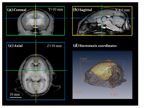
Tissue segmented and population-averaged standard template brain atlas images for the common marmoset were generated by Iriki and Okano in collaboration with colleagues from Keio University School of Medicine, the Central Institute for Experimental Animals in Kawasaki, RIKEN Brain Science Institute, and Fujita Health University School of Health Sciences (Japan) and UCL Institute of Neurology (UK). While the smaller size of marmosets makes them easy to handle it raises other challenges as their brains are typically eleven times smaller than macaques. The researchers used longer data acquisition times to achieve the higher spatial resolution required while allowing for the reduced signal to noise ratio.
In addition the researchers manually applied data analysis techniques to retrieve accurate images of slices through 22 imaged marmoset brains. The researchers noted an area of the corpus callosum that was larger in males than females allowing differences between the sexes to be identified using the brain atlases.
Templates and maps from the study are available at International Neuroinformatics Japan Node website, http://brainatlas.brain.riken.jp/marmoset/index.php?ml_lang=en.
Publication and Affiliation
K. Hikishima 1,3,†, M.M. Quallo 4,5,†, Y. Komaki 1,3, M. Yamada3,6, K. Kawai 3, S. Momoshima 2, H.J. Okano 1, E. Sasaki 3, N. Tamaoki 3, R.N. Lemon 4,5, A. Iriki 4,5*, H. Okano1,* Population-averaged standard template brain atlas for the common marmoset (Callithrix jacchus). Neuromage, 54, 2741-2749, (2011).
- Department of Physiology, Keio University School of Medicine, 35 Shinanomachi, Shinjuku, Tokyo 160-8582, Japan
- Department of Diagnostic Radiology, Keio University School of Medicine, 35 Shinanomachi, Shinjuku, Tokyo 160-8582, Japan
- Central Institute for Experimental Animals, 1430 Nogawa, Miyamae, Kawasaki, Kanagawa, 216-0001, Japan
- Laboratory for Symbolic Cognitive Development, RIKEN Brain Science Institute, 2-1 Hirosawa, Wako, Saitama, 351-0198, Japan
- Sobell Department of Motor Neuroscience and Movement Disorders, UCL Institute of Neurology, London WC1N 3BG, UK
- Faculty of Radiological Technology, Fujita Health University School of Health Sciences, 1-98 Dengakugakubo, Kutsukake-cho, Toyoake-shi, Aichi 470-1192, Japan
*corresponding authors, e-mail address:hidokano@sc.itc.keio.ac.jp
Figure: