


Research Highlights
Vol.14, February 2019
Mapping brain function of monkeys
A novel technology has made it easier to visualize patterns in the brains of naturally behaving non-human primates
Non-human primates (NHP) have very similar social and cognitive processing to humans. Therefore, our knowledge of human behaviour can be advanced by studying the brains of NHPs. Takahiro Kondo, Hideyuki Okano and colleagues at Keio University have developed a new method of imaging neurons in the marmoset brain, a huge advancement in this field.
Traditional imaging studies on NHPs are done using head fixation, in an isolated setting. This renders it impossible to understand cognitive processes of NHPs in a natural setting. The team led by Kondo, developed a method of introducing a miniature lens into the marmoset brain, enabling them to visualize activity without the need for head fixation. For their research, the miniature lens was introduced into the motor cortex; the region responsible for planning and execution of voluntary motor movements.
After carefully mapping the motor cortex, a fluorescent calcium probe was introduced. When neurons are firing, there is influx and efflux of calcium, which can be captured with light emitted from these probes. The prism shaped lens was then fixed in after ensuring it could detect the light signal. Using their technique, up to 80-250 neurons could be captured at a time.
The device was then tested over several movement related tasks the marmosets underwent. It was observed that a subgroup of neurons was active during pulling and climbing movements, activities inherent to the animals. During periods of rest, another set of neurons showed positive calcium signals, as opposed to no activity in the motor cortex. To then study direction-based voluntary movement, the animals were presented with pellets (treats) periodically, located on either their right or left side. It was now seen that more subtypes of neurons were involved, each in the different phases of this task: before reaching for the pellet, during the actual reaching movement, and at the time of retrieval.
In their study, Kondo and co-workers highlighted the role of different neuron subtypes during naturalistic and directional voluntary movement. However, such advanced imaging will also aid in understanding neurological conditions affecting small regions of the brain, such as stroke. They positively conclude that, “This technology will make it possible to dissect large-scale neural circuits during human-relevant behaviour under natural conditions, enabling the study of complex behaviours, including social interaction, fear, and anxiety, and cognitive motor tasks”.
Reference:
Kondo, T. et al. Calcium Transient Dynamics of Neural Ensembles in the Primary Motor Cortex of Naturally Behaving Monkeys. Cell Reports 24, 2191-2195 (2018).
https://doi.org/10.1016/j.celrep.2018.07.057
Figure:
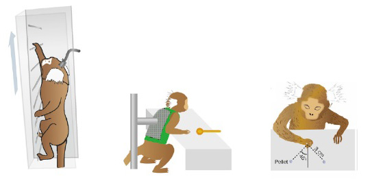
Figure 1. A graphical representation of the ladder climbing (left), lever-pulling (middle) and pellet-retrieval (right) task, the marmosets underwent.